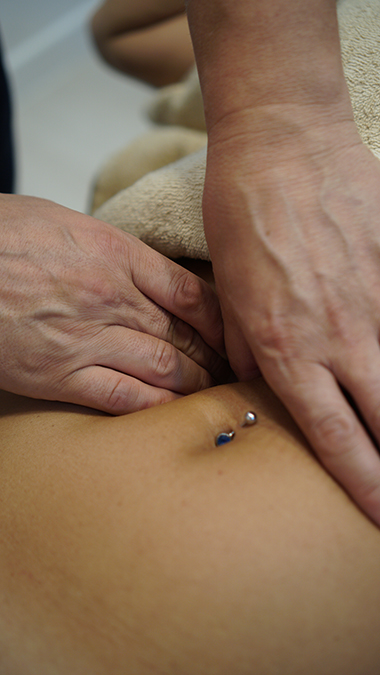
VISCERAL MANIPULATIONS ACCORDING TO BARRAL
The Theory of Visceral ManipulationThe physiology of organ movement
In general, three types of movement are distinguished in the internal organs:
Motricity, Mobility and Motility
Motricity
Motricity refers to the displacement of internal organs relative to the locomotor system, particularly in relation to changes in the spatial position of the spinal column and peripheral joints. For example, when the upper body is bent to the left, the organs on the left side may be subjected to compression or constriction, while the structures on the opposite side will be subjected to tension forces. Similarly, when the body is bent forward, the structures and internal organs at the front will change position in relation to the front and gravity. The peristaltic movements in the small and large intestines of a person who sits continuously will eventually be affected. When both arms are raised to their maximum height, the back section of the spine will bend backwards and the volume of the lungs will expand without taking a deeper breath.Mobility
In visceral manipulation terminology, mobility refers to the movement of both internal organs relative to each other or relative to other fixed structures, particularly the vertebral column or diaphragm. The driving force behind this movement may be various "automatisms" or motricity.Automatisms are involuntary movements of smooth and striated muscles. It is necessary to distinguish between automatisms and periodic movements; examples of automatisms include diaphragmatic breathing, heartbeat, and peristaltic movements of hollow organs.
Motility
It refers to the low-frequency and small-amplitude movements of the internal organs. This movement is the kinetic manifestation of the internal organs, which can only be felt by skilled and experienced hands (low-intensity organ movements).It can be explained as a kind of "inner voice" of the organ, formed as a sort of cellular memory during the migration of organs from their initial positions to their final locations in the womb (during embryonic life).
This movement has two phases:
Expiratory phase – from the outside to the midline
Inspiratory phase – from the midline to the outside
This describes the movement.
Its frequency is 7-8 inh/exp/min
Exhalation phase – from the outside to the midline
Visceral Joint
Motricity, automatism, and mobility cause positional changes in the internal organs. The organs move relative to each other like the joints in the body, moving like a pair of visceral joints between two organs. For example, the liver and kidney, the liver and diaphragm.The surfaces of these structures have a certain degree of slippage over each other, and there are special film-like structures that separate them. For example, the pleura in the lungs, the peritoneum in the intestines, and the meninges in the brain.
The corresponding joint partners are fixed (connected) in certain structures. This is very important because these connection points determine the axes of movement of the organs.
The organs are fixed in various ways.
1. Double-leaf system – the organs are separated by a film or liquid; this structure resembles two glass plates separated by liquid, with a special fluid between them that enables movement.
2. Ligament System - describes special ligaments that are extremely strong and rigid, yet avascular but richly innervated, which hold organs in place against gravity. These structures attach organs to the body.
3. Turgor and anatomical cavity pressure
Turgor defines the maximum expansion and stretching capacity of organs in cavities based on their own elasticity (flexibility). Factors affecting this include the gases within the organs and the pressure of circulation. Cavity pressure is dependent on organ pressure as well as the pressure between organs. The best example of this is stomach hernias. Due to the internal pressure of the abdomen, the stomach migrates into the chest cavity. One of the factors supporting the internal abdominal pressure is the vacuum effect of the diaphragm; therefore, organs close to the diaphragm also maintain their spatial positions thanks to this effect.
4. Mesentery
Mesenteries are structures that attach the outer membrane of the intestines, known as the peritoneum, to the posterior wall of the abdomen: their primary function is to provide structural organisation for circulation rather than support.
5. Omentum
The omenta are folds of the peritoneum and serve no purpose other than to provide interconnections between structures. Their circulatory and nervous functions are more important.
Organ Movement Physiology
All organs move within a specific axis and a specific amplitude (range of motion). Changes in the axis or amplitude alter the physiological movement of the organs.- Local changes are initially asymptomatic, presenting symptoms at a later stage.
- Recurrent local changes
- Intra-abdominal pathologies can affect the entire body via neighbouring organs, vessels, nerves and soft tissue.
Impaired Mobility
The organ loses its mobility partially or completely. The reasons for this are:
Adhesions
Fixations
Vissospasm (muscle tension)
Ptosis (organ sagging)
Amplitude (joint mobility)
Articular restrictions.
In adhesions, motility is restricted while mobility remains normal. Conditions in which the quality of movement is affected are referred to as fixations, and the axis and amplitude of movement change. Causes:
- infections
- inflammations
- surgical procedures
- blunt trauma
Muscle Restrictions (Vissero-spasm)
This refers to non-specific conditions characterised by smooth muscle contractions in hollow organs. As a result, motility is affected, particularly amplitude.
Among the causes of visserospasm:
- inflammation
- vegetative nerves
- allergic reactions
- psychosomatic causes.
Loss of Ligament Elasticity (Ptosis)
Loss of elasticity or tension in ligamentous connections causes organs such as the kidneys, large intestine, bladder, and uterus to sag due to gravity.
Impaired Motility
The amplitude of motility is impaired, range of motion may be impaired in one or two directions, and the rhythm of movement may also be affected. For example, the resting phase of inspiration and expiration may be prolonged. Movement may be arrhythmic or its frequency may change. The reasons for this are:
- general change in vitality
- changes in blood flow
- ptosis
- viscerospasm